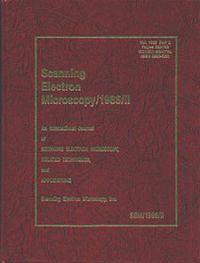
Scanning Electron Microscopy
Volume 1986, Number 2 (1986) Part II
Articles
Low Energy Electron Diffraction with Microscopic Resolution
J. Kirschner, T. Ichinokawa, Y. Ishikawa, M. Kemmochi, N. Ikeda, and Y. Hosokawa
High Resolution Scanning Auger Microscopy of Mineral Surfaces
M. F. Hochella Jr., A. M. Turner, and D. W. Harris
Ion-Induced Auger Emission from Solid Targets
Josette Mischler and Nicole Benazeth
Quantitative Ion Microprobe Analysis of the Rare Earth Elements in Minerals
Ghislaine Crozaz and Ernst Zinner
Charging Effects in Low-Voltage Scanning Electron Microscope Metrology
M. Brunner and R. Schmid
Use of Electron Back Scatter Diffraction Patterns for Determination of Crystal Symmetry Elements
D. J. Dingley and Karim Baba-Kishi
Microtextures of Laterites and Bauxites Capping Deccan Trap Basalts in Western India
D. V. Chitale and N. Güven
Mapping Solid Surfaces with a Raman Microprobe
P. M. Fauchet
Auger Microprobe Temperature Profiles of Contamination Residue on Single Crystal Ferrite Substrates
Christine Bisagni and Nina Veisfeld
Current Filament Formation in Gold Compensated Silicon Pin Diodes
H. Baumann, T. Pioch, H. Dahmen, and D. Jäger
Capacitive Coupling Voltage Contrast
S. Görlich, K. D. Herrmann, W. Reiners, and E. Kubalek
Electron Detectors for Electron-Beam Testing of Ultra Large Scale Integrated Circuits
S. C. J. Garth and D. F. Spicer
Evidence for the use of a Diamond Drill for Bead Making in Sri-Lanka
A. J. Gwinnett and L. Gorelick
Scanning Electron Microscopy in the Evaluation of Consolidation Treatments for Stone
A. E. Charola and R. J. Koestler
Applications of X-Ray Microanalysis to the Study and Conservation of Ancient Glasses
S. Hreglich and M. Verita
The Identification and Characterization of Metal Wrappings in Historic Textiles Using Microscopy and Energy Dispersive X-Ray Spectrometry: Problems Associated with Identification and Characterization
N. Indictor and R. J. Koestler
Weighted Silks Observed Using Energy Dispersive X-Ray Spectrometry
M. Ballard, R. J. Koestler, and N. Indictor
Elemental and Ultrastructural Characteristics of the Egg Capsules of Nautilus Pompilius
R. J. Koestler, E. D. Santoro, and G. Dingerkus
Application of Scanning Electron Microscopy to the Study of Shark Dermal Denticles
G. Dingerkus and R. J. Koestler
Scanning and Transmission Electron Microscopic Study of Recovered Porcine Aortic Valved Conduits
D. J. Allen, I. H. Fentie, J. T. Davis, and Angela Lineen
Intracellular Structure of the Outer Hair Cell of the Organ of Corti
Y. Harada, T. Sakai, N. Tagashira, and M. Suzuki
Scanning Electron Microscopic Observation of the Crista Ampullaris
Y. Harada, M. Takumida, and N. Tagashira
Scanning Electron Microscopy of the Microvascular System in the Inner Ear
Yoshiaki Nakai, Haruhiko Masutani, and Hiromasa Cho
Quantitative Vascular Casting of the Post-Ischemic Hydronephrotic Kidney
Vincent H. Gattone II and Robert D. Sale
The Angioarchitecture of the Lewis Lung Carcinoma in Laboratory Mice (A Light Microscopic and Scanning Electron Microscopic Study)
T. W. Grunt, A. Lametschwandtner, K. Karrer, and O. Staindl
The Characteristic Structural Features of the Blood Vessels of the Lewis Lung Carcinoma (A Light Microscopic and Scanning Electron Microscopic Study)
T. W. Grunt, A. Lametschwandtner, and K. Karrer
Functional Aspects of Renal Glomeruli Based on Scanning Electron Microscopy of Corrosion Casts, with Special Emphasis on Reptiles and Birds
H. Ditrich and H. Splechtna
Immunoarchitecture of the Regenerating Rat Spleen: Effects of Partial Splenectomy and Heterotopic Autotransplantation
Michael C. Dugan, Thomas M. Grogan, Marlys H. Witte, Catherine Rangel, Lynne Richter, Charles L. Witte, and David B. Van Wyck
Structural Features of Isolated, Fractionated Bone Marrow Endothelium Compared to Sinus Endothelium in Situ
Seiji Irie and Mehdi Tavassoli
Ultrastructural Studies of Intercellular Contacts (Junctions) in Bone Marrow. A Review
Ferrell R. Campbell
Adverse Effects of Metals on the Alveolar Part of the Lung
Anne Johansson and Per Camner
Comparative Aspects of Mammalian Spermiogenesis
Leif Plöen and Jean-Luc Courtens
Cells from Xenopus laevis Gastrulae Adhere to Fibronectin-Sepharose Beads and Other Lectin Coated Beads
Kurt E. Johnson and Michael H. Silver
Ultrasonic Microdissection of Immature Intermediate Human Placental Villi as Studied by Scanning Electron Microscopy
Gregory J. Highison and F. Donald Tibbitts
Correlative Scanning Electron Microscopy in the Study of Human Gastric Mucosa
F. Bonvicini, M. C. Maltarello, P. Versura, D. Bianchi, G. Gasbarrini, and R. Laschi
Preparation Methods for Quantitative Electron Probe X-Ray Microanalysis of Rat Exocrine Pancreas: A Review
N. Roos and T. Barnard
The Use of a Line Scan Ratemeter for the X-Ray Microanalytic Evaluation of Membrane-Bound Histochemical Endproducts
Imre Zs.-Nagy and Valéria Zs.-Nagy
Intrauterine Device (IUD) Associated Pathology: A Review of Pathogenic Mechanisms
Waldemar A. Schmidt and Karmen L. Schmidt
Urolithiasis in a Patient Ingesting Pure Silica: A Scanning Electron Microscopy Study
D. B. Leusmann, J. Pohl, and G. Kleinhans
Histochemistry of Colloidal Iron Stained Crystal Associated Material in Urinary Stones and Experimentally Induced Intrarenal Deposits in Rats
Saeed R. Khan and Raymond L. Hackett
Lack of Regional Surface Differences in Mouse Bladder Urothelium: A Scanning Electron Microscopic Study
Kari Feren and Jon B. Reitan
Scanning Electron Microscopy of the Irradiated Mouse Bladder Urothelium
Jon B. Reitan and Kari Feren
Statoconia Formation in Molluscan Statocysts
Michael L. Wiederhold, Christine E. Sheridan, and Nancy K. R. Smith


