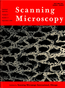
Scanning Microscopy
Volume 5, Number 3 (1991)
Articles
Low Voltage Scanning Electron Microscopy Cathodoluminescence Observations of Gallium Arsenide
S. Myhajlenko
Digitized Cathodoluminescence Imaging of Minerals
Ian M. Steele
Enhancing the Laser Scanning Confocal Microscopic Visualization of Lucifer Yellow Filled Cells in Whole-Mounted Tissue
David L. Becker, Joanna Dekkers, Roberto Navarrete, Colin R. Green, and Jeremy E. Cook
Scanning Tunneling Microscope Images of Adenine and Thymine at Atomic Resolution
M. J. Allen, M. Balooch, S. Subbiah, R. J. Tench, W. Siekhaus, and R. Balhorn
High-Resolution, Real-Space Imaging of Conformational Structures of Poly-L-Proline Helixes
N. J. Zheng, G. Rau, C. F. Hazlewood, and C. Rau
Band Positions Used for On-Line Crystallographic Orientation Determination from Electron Back Scattering Patterns
N. H. Schmidt, J. B. Bilde-Sørensen, and D. Juul Jensen
Scanning Electron Microscopy to Establish the Marble Weathering Mechanism in the Alhambra of Granada (Spain)
M. A. Bello, L. Martín, and A. Martín
Prerequisites of High Resolution Scanning Electron Microscopy
René Hermann and Martin Müller
Scanning Electron Microscopy of High-Modulus Polyethylene Fibres
N. H. Ladizesky and M. K. M. Pang
The Measurement of Aluminum Surface Diffusion on Si, SiO2, and Si3N4 by Scanning Auger Microscopy
L. L. Levenson, A. B. Swartzlander, A. Yahashi, H. Usui, and I. Yamada
Influence of Ageing, pH and Various Additives on Crystal Formation in Artificial Urine
A. L. Rodgers and M. A. E. Wandt
Retention of Calcium Oxalate Crystals in Renal Tubules
Saeed R. Khan and Raymond L. Hackett
Heterogeneity of Crystals Attached to the Human Enamel and Cementum Surfaces After Calculus Removal In Vitro
T. Kodaka, K. Debari, and M. Yamada
Morphological Study of Calcospherites in Rat and Rabbit Incisor Dentin
H. Mishima, T. Sakae, and Y. Kozawa
In Vitro and In Vivo Replication for Scanning Electron Microscopy of the Cervical Region of Human Teeth
Joan Bevenius and Kjell Hultenby
Localisation of Putative Mechanoelectrical Transducer Channels in Cochlear Hair Cells by Immunoelectron Microscopy
Carole M. Hackney, David N. Furness, and Dale J. Benos
Three Dimensional Intracellular Structure of the Cochlea Using the A-O-D-O Method
A. Nagasawa, R. V. Harrison, R. J. Mount, and Y. Harada
The Vestibular Epithelia in Experimental Hydrops
K. C. Horner and S. Rydmarker
Beam Sensitivity of Globoid Crystals within Seed Protein Bodies and Commercially Prepared Phytates During X-Ray Microanalysis
Irene Ockenden and John N. A. Lott
Seed Coat Surface Patterns and Structures of Oxytropis riparia, Oxytropis campestris, Medicago sativa, and Astragalus cicer
D. J. Solum and R. H. Lockerman
Effect of a Platinum Chemotherapy Drug on Intracellular Elements During the Cell Cycle, Using X-Ray Microanalysis
P. G. Edwards, M. D. Kendall, and I. W. Morris
The Vascularization of the Digestive Tract Studied by Scanning Electron Microscopy with Special Emphasis on the Teeth, Esophagus, Stomach, Small and Large Intestine, Pancreas, and Liver
S. Aharinejad, A. Lametschwandtner, P. Franz, and W. Firbas
Hereditary Hair Changes Revealed by Analysis of Single Hair Fibres by Scanning Electron Microscopy
B. Forslind, M. K. Andersson, and E. Alsterborg
Proton Induced X-Ray Emission Analysis of Biological Specimens - Past and Future
B. Forslind, K. G. Malmqvist, and J. Pallon
Elemental Analysis and Fine Structure of Mitochondrial Granules in Growth Plate Chondrocytes Studied by Electron Energy Loss Spectroscopy and Energy Dispersive X-Ray Microanalysis
Joanna Wroblewski, Romuald Wróblewski, Claudie Mory, and Christian Colliex


